The story of Maurya
The arrival of our son, Maurya on July 11, 2012 was a watershed moment for us When our son, Maurya was one year old, we (my wife Yash and I) noticed some “delays” in Maurya’s developmental milestones. The doctors did not see any red flags; they said that every child was different and not to worry. We were not too concerned then and enjoying our happy moments as a family. Our worries began to unfold when Maurya was eighteen months old and he reached the universal range for not meeting the walking milestone. We contacted Maurya’s pediatrician and they suggested physical therapy once a week. A few months passed with no signs of improvement.
In March 2014, Maurya was still not walking and we began to see “red flags”. Maurya was twenty months old at this point. My wife contacted Early Intervention at Illinois State and they started their evaluation. The Early Intervention team concluded that Maurya had a developmental delay with both his walking and his speech; his speech delay was on the mild side. We started physical therapy and speech therapy, physical therapy one day a week, and speech therapy for one day a week. Supra-Malleolar Orthosis (SMO) was prescribed by physical therapists. SMOs are the inserts within the shoe that help stabilize the ankle and helps with walking.
Physical therapists later prescribed a reverse walker for Maurya. Reverse walkers (also called posture control walkers) are positioned behind Maurya to enable a more upright posture. Maurya takes a step and then pulls the walker up behind him before taking another step. He slowly, but progressively, started getting adapted and has been using this walker since June 2014.
In September 2014 (Maurya was twenty-six months old), we met with Dr. Karen Manning, a local neurologist at DuPage Medical Group. Dr. Manning then ordered a brain MRI for Maurya. The MRI showed no abnormalities. As part of the Early Intervention Program, the coordinators scheduled us to meet a physiatrist at the MarianJoy Rehabilitation Centre in Wheaton, Illinois and in October 2014, we met Dr. Mary Keen. She made an evaluation and concluded that Maurya (now twenty-seven months old) had Cerebral Palsy. Cerebral Palsy is an umbrella of permanent movement disorders that appear in early childhood. Dr. Keen suggested that we continue the physical and speech therapies. That November, per Dr. Keen’s suggestion, we gave Maurya some ankle support through Ankle-Foot Orthosis or AFO. AFO is support intended to control the position and motion of the ankle and compensate for weakness. Physical therapies were then administered to Maurya three times a week. We continued those therapies at Early Intervention as well as at a separate private pediatric facility.
We began to question the Cerebral Palsy diagnosis because of his normal brain MRI. We were told that in many cases, even with a normal brain MRI, there could be hidden abnormalities. The brain is a complex organ. The ability to investigate brain functions in their entirety with an MRI for accurate diagnoses remains limited. All his symptoms were consistent with Cerebral Palsy and so, we were convinced of the accuracy of the Cerebral Palsy diagnosis.
Maurya’s physical, occupational and speech therapies continued in 2015. We continued the therapies with Illinois State’s Early Intervention until Maurya’s third birthday in July 2015. Early Intervention was only available until a child reaches three years of age. We then continued the therapies in a private clinic. Then, on the advice of the physical therapist, we tried equine therapy (horseback riding or hippotherapy) for strength building. Maurya did this for about three months (once a week), with no major improvement. In late 2015, we enrolled him in swimming lessons until that December, and he did well. We also enrolled Maurya in an intensive ten-week TheraSuit program at Easter Seals in Villa Park Illinois.
As part of our ongoing research to pin down the diagnosis, we had consulted our neurologist again in early 2016 to inquire about further options. The neurologist then suggested a spinal MRI. The spine MRI showed two syrinxes in the spinal cord, cervical and thoracic sections (a syrinx is a fluid-filled cavity within the spinal cord). The cervical and thoracic sections are the top and middle sections of the spine. This yielded a new diagnosis- Syringomyelia. The neurologist suggested that we see a neurosurgeon and referred us to Dr. David Frim from the University of Chicago.
Dr. Frim saw Maurya in April 2016. He concluded that his condition had no bearing on his intelligence and that there was no correlation between the syrinx in his spine and his inability to stand and walk. Further, he did not have an answer as to why Maurya was unable to stand and walk. Dr. Frim suggested we continue all three of his therapies- Physical, Occupational, and Speech. In August of 2016, we followed up with Dr. Frim for another MRI. The size of the syrinx was unchanged.
In September 2016, I took Maurya to Gillette Children’s Specialty Healthcare in St. Paul, Minnesota. They arranged a team of doctors and surprisingly, Dr. Patrick Graupman, a neurosurgeon, stated that Maurya had a tethered spinal cord and suggested surgery. This was yet another new twist. Maurya had a new diagnosis: Tethered cord. Tethered spinal cord condition is a neurologic disorder caused by tissue attachments that limit the movement of the spinal cord within the spinal canal. The spinal cord normally floats free inside the spinal canal.
For second opinion, my wife met with Dr. Anandh Balasubramaniam, a neurosurgeon at Yashoda Hospitals, Secunderabad, India bringing with her the existing MRIs. Dr. Balasubramaniam could not confirm the tethered cord diagnosis, as there had been no lumbar MRI of the spine to review. The lumbar section is the bottom section of the spine. At this point, we wanted a third opinion to confirm or negate the diagnosis. In October 2016, we met with neurosurgeon Dr. Lorenzo Munoz of Rush University Medical Center in Chicago. We only had Maurya’s thoracic and cervical spine MRIs, so Dr. Munoz also suggested a lumbar MRI to rule out a tethered spinal cord. They performed the MRI and ruled out the tethered cord. We shared the lumbar MRI with Dr. Anandh Balasubramaniam in Secunderabad, India and he ruled out tethered cord also.
We met Dr. Munoz again in December 2016. He wanted to perform a Cerebrospinal Fluid (CSF) flow study to examine the uniformity of CSF fluid. The study had negative results. He then repeated the MRI for the spine (cervical and thoracic regions). There had been no change since the August 2016 MRI at the University of Chicago.
While this was happening, we also enrolled Maurya in a neurofeedback program for speech improvement. Maurya was verbal and communicative but struggled to frame sentences and had great difficulty narrating a story. He followed instructions well and always responded in one or two sentences. One of the neurologists mentioned neurofeedback and suggested we try it out. Neurofeedback provides immediate feedback from a computer-based program that assesses the patient’s brainwave activity while hooked to an EEG (Electroencephalogram). The program teaches the central nervous system to reorganize and regulate the brainwave frequencies.
We did neurofeedback twice a week starting in January and continuing until August of 2016. This resulted in some progress in his speech. Maurya was able to tell short stories although his voice was feeble, and his pace was a little bit slow.
In August 2017, we followed up with Dr. Frim, a one-year follow-up and we repeated the MRI. Dr. Frim confirmed an increase in the size of the syrinx in the thoracic region and suggested spine surgery that would involve the draining of his syrinx through a subarachnoid shunt. He surmised a relationship to Maurya’s decreasing gross motor functions. A syrinx to subarachnoid shunt is used to restore the natural flow of cerebrospinal fluid.
On Sep 25, 2017, Dr. Frim performed the thoracic region surgery at the University of Chicago; the surgery was done in the thoracic region of the spine. The cervical syrinx was untouched as planned. Maurya was also prescribed rehabilitation for three weeks to regain his strength. We followed up with Dr. Frim again in January of 2018 and repeated the MRI. The spine now was crystal clear with no syrinx, but it never solved the original problem of Maurya’s inability to stand and walk independently. Syringomyelia as the cause of Maurya’s condition was ruled out.
Physical therapy, occupational therapy, and swimming resumed for Maurya for all of 2018.
We thought that having an orthopedic specialist’s opinion would be worthwhile; we previously had not considered the orthopedic dimension. We made an appointment with Dr. Altiok Haluk, an orthopedic surgeon from Shriners Hospital in Chicago in October 2018. Dr. Altiok suggested that Maurya needed bilateral hip surgery (Femur Varus Osteotomy) for both hips. When a hip starts to move out of joint, the ball which is at the head of the femur is no longer contained properly within the socket. A Varus Osteotomy is performed to place the ball back into the socket. Dr. Altiok also suggested an additional Acetabuloplasty for the right hip. Acetabuloplasty involves taking away the area of over coverage on the Acetabular side (socket of the hip bone) to create space allowing normal motion of the hip.
Maurya’s regular Physiatrist is Dr. Larissa Pavone at MarianJoy. We take Maurya to her regularly for his orthotics, walker, wheelchair, and his developmental appointments. She referred to Dr. Nancy L. Kuntz at Lurie Children’s Hospital of Chicago. Dr. Kuntz specializes in neuromuscular disorders in children with special emphasis on neuroimmunology and autonomic disorders. We met with Dr. Kuntz in October 2018 to hear her opinion. Dr. Kuntz suggested a lumbar puncture of Maurya’s spine for fluid study and dystonia. Dystonia is a movement disorder. It can affect one or more parts of the body, and sometimes the entire body. The condition can be mild or severe. However, this test was negative, so she then suggested seeing a geneticist for advanced genetic testing. We already had done basic genetic testing for Maurya, with no definitive findings.
During February and March of 2019, we learned that serial casting could help Maurya, so we did that for close to eight weeks. Serial casting is the process of putting on and removing a cast on a leg every five to six days for several weeks. The goal is to stretch the muscles to allow for better movement and range of motion over. This was a total waste of time and effort as his unexpected bi-lateral hip surgery surfaced in May 2019 which reset Maurya’s walking.
In late March of 2019, per Dr. Kuntz’s suggestion. we met Dr. Darrel Waggoner, a Clinical geneticist for genome testing (Advanced genetic testing) from the University of Chicago. Meanwhile. we have decided to go ahead with Dr. Altiok’s suggested hip surgery at Shriners Hospital in Chicago on May 20th, 2019.
We heard back after multiple weeks of testing from Dr. Darrel Waggoner, the Clinical geneticist for genome testing (Advanced genetic testing) from the University of Chicago. On May 23, 2019, after years of struggle, the answers to our unknowns were finally unveiled. The geneticist confirmed that there was an indication of the SPAST4 gene in Maurya’s bloodwork. SPAST4, commonly known as SPG4 (spastic paraplegia type 4) is part of a group of genetic disorders known as hereditary spastic paraplegias (HSP). These disorders are characterized by progressive muscle stiffness (spasticity) and the development of paralysis of the lower limbs (paraplegia). As this disorder is usually hereditary, Yash and I were also tested to check if we were possible carriers.
The results showed that we were not the carriers, so Maurya now fell in a low probability range for these disorders. Maurya happened to be in the category of de-novo mutation. A de-novo mutation is a genetic alteration that is present for the first time in one family member as a result of a variant (or mutation) in a germ cell (egg or sperm). HSP is characterized by insidiously progressive bilateral lower-limb gait spasticity. What we came to know in our research is that it is often misdiagnosed.
Maurya had a break in physical and Occupational therapies from March through July of 2020 due to pandemic. I had couple of curative surgeries and during this entire period, Maurya did not receive PT/OT. Maurya mobility gradually decreased during this period and continued its course in 2021 and in 2022 and Maurya is now completely wheelchair bound. He struggles very hard to take few steps using his gait trainer with Physical Therapist’s support in a private physical therapy set up but very little to no improvement. Since January of 2022, Maurya is having slow, slurred speech issues with feeble voice. He is better some days but overall, we are noticing the downward spiral in speech since 2022. To this day (February 14, 2023), we are continuing with Physical, Occupational and Speech therapies.
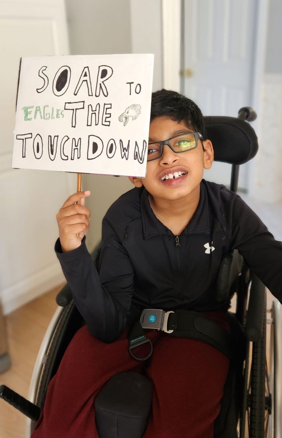
While all of this is happening to Maurya’s physicality, he is still a happy kid with an infectious smile and with lot of motivation and interest in academics- Math, English. He loves to travel, have a great repository of animals, absolutely love NBA and NFL games, and Maurya plays wheelchair basketball and plays piano well. Last but not the least, he is a great Lego builder and has built numerous architectural Lego structures.